SIMILAR EFFICACY TO LUCENTIS IN THE TREATMENT OF nAMD1,2

A Phase 3 Clinical Trial Comparing BYOOVIZ and Lucentis1,2

A Phase 3 Clinical Trial Comparing BYOOVIZ and Lucentis1,2 A Phase 3 Clinical Trial Comparing BYOOVIZ and Lucentis1,2

*

The endpoints were chosen in consultation with appropriate regulatory bodies. BCVA was the selected primary endpoint for FDA approval.1

Active CNV indicated presence of leakage and intra- or subretinal fluid and was confirmed by central reading center during screening.1

Primary Endpoint: Difference of LS Mean Change in BCVA Between BYOOVIZ and Lucentis at Week 81

Primary Endpoint: Difference of LS Mean Change in BCVA Between BYOOVIZ and Lucentis at Week 81

Difference in LS mean change from baseline in BCVA at Week 8 in the full analysis set (FAS) (BYOOVIZ − Lucentis); whiskers represent the 90% CI that is contained within the predefined equivalence margins of −3 to 3 letters, represented by the dashed lines.

n=351 (BYOOVIZ), n=353 (Lucentis). The FAS included all randomized participants, excluding 1 inadvertently randomized participant who did not receive study drug.1

  • At Week 8, visual acuity for BYOOVIZ was within the predefined equivalence margins compared with Lucentis1
  • Adjusted treatment difference between groups was −0.8 letters (90% CI: −1.8 to 0.2 letters)1

Primary Endpoint: Difference of LS Mean Change in CST Between BYOOVIZ and Lucentis at Week 41

Primary Endpoint: Difference of LS Mean Change in CST Between BYOOVIZ and Lucentis at Week 41

Difference in LS mean change from baseline in CST at Week 4 in per-protocol set (PPS) (BYOOVIZ – Lucentis); whiskers represent the 95% CI that is contained within the predefined equivalence margin of −36 to 36 μm, represented by the dashed lines.

n=342 (BYOOVIZ), n=338 (Lucentis).

  • At Week 4, CST for BYOOVIZ was within the predefined equivalence margins compared with Lucentis1
  • Adjusted treatment difference between groups was –8.4 μm (95% CI: –19.4 to 2.7)1

1-Year/ Study: Change From Baseline in BCVA at Each Time Point Through Week 52 in the FAS2*

1-Year/ Study: Change From Baseline in BCVA at Each Time Point Through Week 52 in the FAS2*
TAP TO EXPAND TIME POINTS THROUGH WEEK 52
Week | BYOOVIZ, n | Lucentis, n

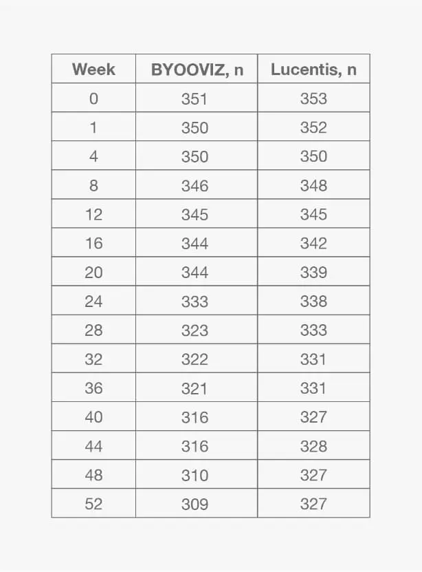
Mean (SD) change from baseline in BCVA for participants who completed Week 52 of the study: BYOOVIZ (n=309), 9.7 (11.4) letters; Lucentis (n=327), 10.4 (11.5) letters. Circles and squares represent mean and error bars represent SE at each time point.

*For additional information and all other secondary endpoints, please refer to Bressler NM, et al., 2021.2
  • Change from baseline in BCVA was 9.8 letters for BYOOVIZ and 10.4 letters for Lucentis at Week 52 (adjusted treatment difference [SE]: −0.6 [0.9]; 90% CI: −2.1 to –0.9 letters)2
  • The proportion of participants who lost <15 letters and who gained >15 letters was comparable between treatment groups at all time points2

1-Year Study: Change From Baseline in CST at Each Time Point Through Week 52 in the FAS2

1-Year Study: Change From Baseline in CST at Each Time Point Through Week 52 in the FAS2
TAP TO EXPAND TIME POINTS THROUGH WEEK 52
Week | BYOOVIZ, n | Lucentis, n
Mean (SD) change from baseline in CST for participants who completed Week 52 of the study: BYOOVIZ (n=308), –133.6 (103.9) μm; Lucentis (n=327), –128.4 (116.1 μm). Circles and squares represent mean and error bars represent SE at each time point.
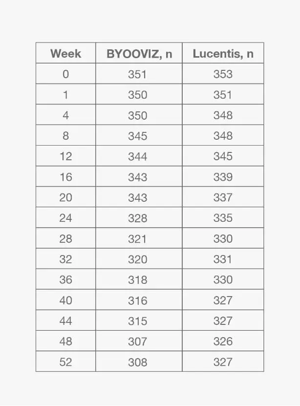
  • Change from baseline in CST was −140.0 μm for BYOOVIZ and −125.1 μm for Lucentis at Week 52 (adjusted treatment difference [SE]: −14.9 [5.3]; 95% CI: –25.3 to –4.5)2
  • The change from baseline in CST was comparable between treatment groups at all time points up to Week 522
BCVA=best corrected visual acuity (ETDRS letter score); CI=confidence interval; CNV=choroidal neovascularization; CRLT=central retinal lesion thickness; CST=central subfield thickness; DA=disc area; ETDRS=Early Treatment Diabetic Retinopathy Study; ITV=intravitreal; LS=least squares; nAMD=neovascular (wet) age-related macular degeneration; SD=standard deviation; SE=standard error.
Phase 3 data support the biosimilarity of BYOOVIZ to Lucentis1,2
Indication and Important
Safety Information
INDICATIONS
BYOOVIZ® (ranibizumab-nuna), a vascular endothelial growth factor (VEGF) inhibitor, is indicated for the treatment of patients with:
  • Neovascular (Wet) Age-Related Macular Degeneration (AMD)
  • Macular Edema Following Retinal Vein Occlusion (RVO)
  • Myopic Choroidal Neovascularization (mCNV)
IMPORTANT SAFETY INFORMATION
CONTRAINDICATIONS
  • BYOOVIZ is contraindicated in patients with ocular or periocular infections or known hypersensitivity to ranibizumab products or any of the excipients in BYOOVIZ
  • Hypersensitivity reactions may manifest as severe intraocular inflammation
WARNINGS AND PRECAUTIONS Endophthalmitis and Retinal Detachments
  • Intravitreal injections, including those with ranibizumab products, have been associated with endophthalmitis, retinal detachments, and iatrogenic traumatic cataract. Proper aseptic injection technique should always be used when administering BYOOVIZ. In addition, patients should be monitored following the injection to permit early treatment, should an infection occur
Increases in Intraocular Pressure
  • Increases in intraocular pressure have been noted both pre-injection and post-injection (at 60 minutes) while being treated with ranibizumab products. Monitor intraocular pressure prior to and following intravitreal injection with BYOOVIZ and manage appropriately
Thromboembolic Events
  • Although there was a low rate of arterial thromboembolic events (ATEs) observed in the ranibizumab clinical trials, there is a potential risk of ATEs following intravitreal use of VEGF inhibitors. ATEs are defined as nonfatal stroke, nonfatal myocardial infarction, or vascular death (including deaths of unknown cause)
  • Neovascular (Wet) Age-Related Macular Degeneration
    • The ATE rate in the three controlled neovascular AMD studies (AMD-1, AMD-2, AMD-3) during the first year was 1.9% (17 of 874) in the combined group of patients treated with 0.3 mg or 0.5 mg ranibizumab compared with 1.1% (5 of 441) in patients from the control arms. In the second year of Studies AMD-1 and AMD-2, the ATE rate was 2.6% (19 of 721) in the combined group of ranibizumab-treated patients compared with 2.9% (10 of 344) in patients from the control arms. In Study AMD-4, the ATE rates observed in the 0.5 mg arms during the first and second year were similar to rates observed in Studies AMD-1, AMD-2, and AMD-3

      In a pooled analysis of 2-year controlled studies [AMD-1, AMD-2, and a study of ranibizumab used adjunctively with verteporfin photodynamic therapy (PDT)], the stroke rate (including both ischemic and hemorrhagic stroke) was 2.7% (13 of 484) in patients treated with 0.5 mg of ranibizumab compared to 1.1% (5 of 435) in patients in the control arms [odds ratio 2.2 (95% confidence interval; 0.8-7.1)]

  • Macular Edema Following Retinal Vein Occlusion
    • The ATE rate in the two controlled RVO studies during the first 6 months was 0.8% in both the ranibizumab and control arms of the studies (4 of 525 in the combined group of patients treated with 0.3 mg or 0.5 mg ranibizumab and 2 of 260 in the control arms). The stroke rate was 0.2% (1 of 525) in the combined group of ranibizumab-treated patients compared to 0.4% (1 of 260) in the control arms
Retinal Vasculitis With or Without Occlusion
  • Retinal vasculitis with or without occlusion, typically in the presence of preexisting intraocular inflammation or post-treatment with other intravitreal agents, have been reported with the use of ranibizumab products. Discontinue treatment with BYOOVIZ in patients who develop these events. Patients should be instructed to report any change in vision without delay

ADVERSE REACTIONS

  • Serious adverse reactions related to the injection procedure have occurred in <0.1% of intravitreal injections, including endophthalmitis, rhegmatogenous retinal detachment, and iatrogenic traumatic cataract
  • The most common adverse reactions (reported more frequently in ranibizumab-treated subjects than control subjects) are conjunctival hemorrhage, eye pain, vitreous floaters, and increased intraocular pressure. The most common non-ocular adverse reactions included nasopharyngitis, anemia, nausea, and cough
  • As with all therapeutic proteins, there is potential for immunogenicity. The clinical significance of immunoreactivity to ranibizumab products is unclear at this time

You are encouraged to report suspected adverse reactions to the FDA. Visit www.fda.gov/medwatch, or call 1-800-FDA-1088.

Please see full Prescribing Information.